Actualités

24 avril 2024
Programme NeuroFOCUS | Brisez l’isolement, restez actives et actifs et optimisez vos capacités!


23 avril 2024
La Feuille de route gouvernementale en économie circulaire au Québec saluée par l’ÉTS, le CERIEC, l’Institut AdapT et le RRECQ
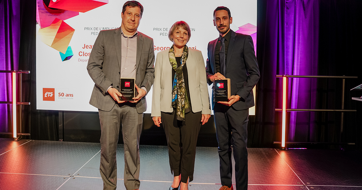
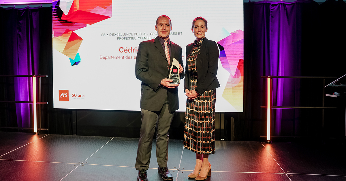
23 avril 2024
Gala d'excellence : Jean-Sébastien Closson-Duquette et Georges Ghazi reconnus par le Décanat des études
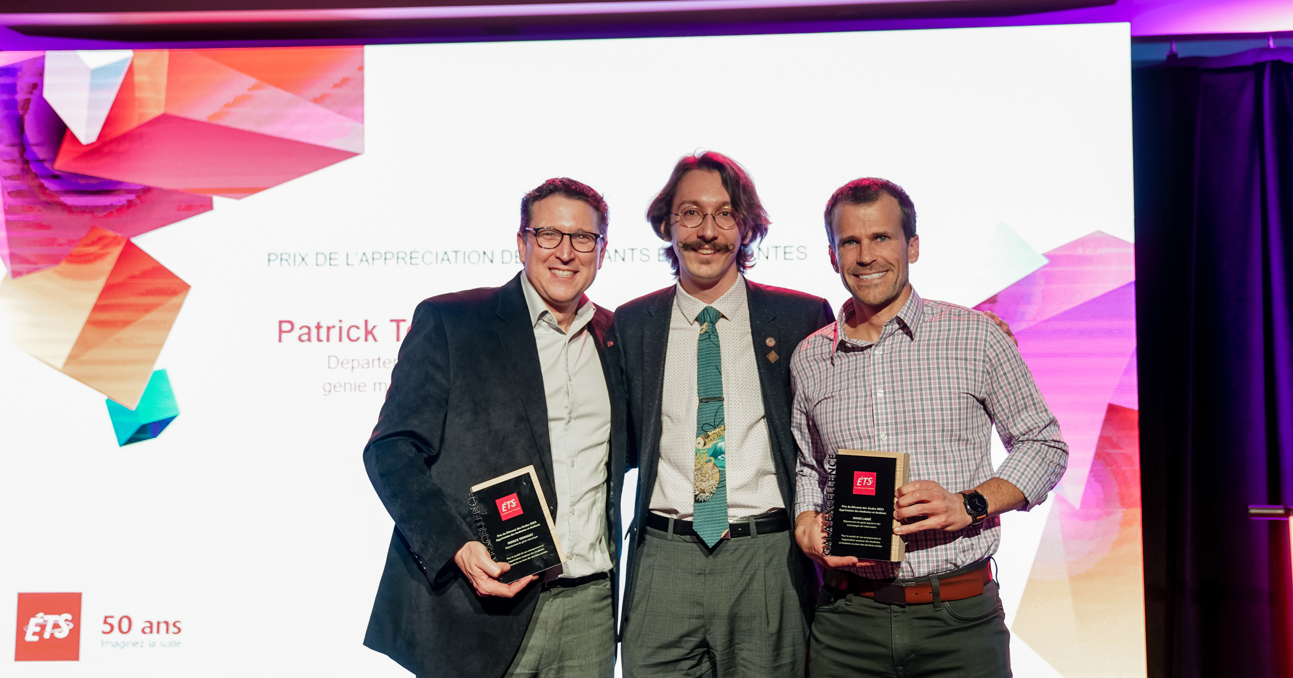
22 avril 2024
Gala d’excellence : Patrick Terriault et David Labbé reçoivent le Prix de l’appréciation des étudiants

17 avril 2024
Fin de session et recherche de stage
1
Vous êtes actuellement sur cette page
2
Aller à la page : 2
3
Aller à la page : 3
...
52
Aller à la page : 52
Aller à la page suivante
Explorez votre avenir
universitaire